
Biophotonics, the interaction between light and biological material, serves as the basis for various medical applications. The following research efforts and technologies highlight some ways in which medical lasers and biophotonics continue to contribute to the refinement of diagnostics and medicine.
Medical Lasers in Diagnosis

Scientists at the Fraunhofer Institute for Photonic Microsystems IPMS in Dresden developed a laser-based sensor that incorporates an 8-mm-diameter micro-electro-mechanical system (MEMS) microscope head. The microscope optically resolves and magnifies tissue cells measuring just 10–20 micrometers. When installed at the tip of an endoscope, it may be used for in vivo cancer diagnosis, potentially eliminating the need for a biopsy. The laser conducts a transmitting fiber to the microscanner mirror fitted in the tip of the endoscope, deflecting the laser beam and illuminating the tissue in question. A glass-fiber bundle located in the tip of the endoscope transmits the reflected light to the external sensor, which then receives a signal containing the image information. A detector measures the position of the scanner mirror, indicating which area of the scene is being illuminated at a specific point in time. A two-dimensional image can be completely reconstructed by combining the position and image sensor signals.
Optical Biopsy for Colonoscopy
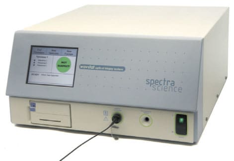
According to the National Cancer Institute, colorectal cancer is the third most commonly diagnosed cancer for both men and women in the U.S. However, between 1984 and 2004, the incidence of colorectal cancer has declined by nearly 26 percent, likely due to increased colorectal cancer screening, which allows physicians to detect and remove colorectal polyps (irregular clumps of cells that form on the lining of the colon) before they potentially become cancerous. The WavSTAT® Optical Biopsy System from SpectraScience (spectrascience.com) in San Diego, CA is designed to be used in conjunction with colonoscopy to allow physicians to better evaluate smaller polyps. Typically, larger polyps are removed and sent to the lab for a biopsy, while smaller/flat polyps are evaluated through visual inspection. The optical biopsy system makes it easier for physicians to take a closer look at smaller or flat polyps by immediately taking a tissue biopsy in the event that an area demonstrates a suspect reaction to light.
A type of technique called low-coherence enhanced backscattering spectroscopy (LEBS) may also be beneficial as an inexpensive, non-invasive test for routine colon cancer screening, an attractive alternative to a full-fledged colonoscopy. Northwestern University researchers developed a screening technique that utilizes this method to analyze tissue samples taken from the base of the rectum. Light shines on the tissue and scatters; some of that light bounces back to sensors in the probe. A computer analyzes the pattern of light scattering and looks for a sign of carcinogenesis in the nanoarchitecture of the cells. The researchers found that LEBS could detect the presence of growths elsewhere in the colon, even though it only analyzed tissue from the base of the rectum. Researchers call this the “field effect,” which means that tissue that looks normal and is located far away from the lesion will still demonstrate molecular changes in LEBS analysis. The study examined tissue from 219 patients; results indicated that such changes could be detected accurately by using LEBS, and that LEBS could be an effective basis for a minimally invasive colon cancer screening technique. Researchers are currently working toward the development of a compatible fiber optic probe.
Medical Lasers in Cytoscopy
The Photodynamic Diagnostic D-Light C (PDD) System from Karl Storz Endoscopy-America (karlstorz.com) in El Segundo, CA was recently approved by the FDA as an optional accessory for white light cystoscopy, a test used to detect cancer of the bladder. The system consists of a D-light C light source, rigid PDD telescopes, fluid light cables, a PDD camera head, and PDD camera control units. The D-light C unit is a 300-watt short arc Xenon light source with two modes of operation: the white light (WL) mode and the PDD mode. The WL mode emits light in the visible spectrum ranging from 390–790 nm and is used for illumination of the bladder during a cystoscopy. The PDD mode emits light in the blue portion of the visible spectrum, from 360–450 nm, and is used to induce and view fluorescence in the bladder. The system interacts with a diagnostic imaging drug called Cysview®. Light is transmitted through a fluid light cable connected to an endoscope, and blue light is used to excite the drug that has collected in cancerous tissue. Tumorous areas fluoresce in red, while normal tissue exhibits a blue color.
Time-Resolved Fluorescence Spectroscopy
Breast Cancer Diagnosis
A handheld laser scanner developed at the UC Irvine Beckman Laser Institute may lead to improved breast cancer diagnosis. The scanner generates a spectral “fingerprint” of each patient in order to determine whether or not breast tumors require more aggressive treatment. Unlike a mammogram, this device can provide detailed metabolic measurements of hemoglobin, fat and water content, tumor oxygen consumption, and tissue density. These measurements also allow physicians to see how the chemotherapy is affecting the cancer, and allow them to adjust treatments accordingly. The scanning method would particularly improve detection in younger women, whose denser breast tissue is less sensitive to mammography. The device is currently being evaluated in a cross-institutional clinical study by Beckman Laser Institute, University of Pennsylvania, Dartmouth College, UC San Francisco, and Massachusetts General Hospital.
Treating Irregular Heartbeats With Lasers

Photochemical Tissue Bonding
An innovative light-activated nanosuture technology known as photochemical tissue bonding (PTB) could be a useful for treating battlefield injuries. The research, managed by Air Force Research Laboratory and funded by the Office of the Secretary of Defense, is taking place at Harvard University's Wellman Center for Photomedicine and Massachusetts General Hospital. PTB could be used not only for closing skin wounds, but also for more delicate procedures, such as reconnecting severed peripheral nerves, blood vessels, tendons, and incisions in the cornea.
The technique works by applying an FDA-approved ophthalmological dye called Rose Bengal to the damaged tissue surface. Clinical lasers emitting CW green light (532 nm) are then applied to the area. When the dye reacts with the light, collagen in the affected tissue area is reconnected to form a watertight seal. Bench-level lab experiments indicate that this technology reduces near-term inflammation and minimizes scarring, compared to conventional sutures.
PTB technology is still being tested and refined in clinical trials. Although it currently takes three minutes to close a wound using this method, researchers hope to whittle the process down to less than 30 seconds.

