Researchers at the University of British Columbia, Vancouver, BC, Canada, have developed a new magnetic resonance imaging (MRI) technique that detects signs of multiple sclerosis (MS) in finer detail than ever before. This, they say, provides a more powerful tool to evaluate new treatments.
The technique analyzes the frequency of electro-magnetic waves collected by an MRI scanner, instead of the size of those waves. Although analyzing the number of waves per second had long been considered a more sensitive way of detecting changes in tissue structure, the math needed to create usable images had proved daunting.
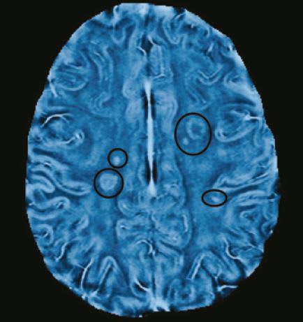
MS occurs when a person’s immune cells attack the layer of myelin that protects nerve fibers. The myelin breakdown interferes with the electrical signals transmitted between neurons, leading to a range of symptoms, including numbness or weakness, vision loss, tremors, dizziness, and fatigue.
The researchers applied their method to 20 MS patients, who were scanned once a month for six months using both conventional MRI and the new frequency-based method.
Once lesions in the myelin appeared in conventional MRI scans, the scientists went back to earlier frequency-based images of those patients. And, by examining the precise areas of those lesions, they found frequency changes, which indicate tissue damage, at least two months before any sign of damage appeared on conventional scans.