Diabetic retinopathy resulting from long-term diabetes mellitus is one of the common diseases that leads to choroidal neovascularization (CNV), a leading cause of blindness. Among the currently available treatment methods, a laser can be used to photocoagulate the diseased areas. Several thousand laser shots are usually required during such treatment. Special care must be taken to avoid hitting the blood vessel tree, the macula, the optic disk, and the region among them. For a single eye, this procedure requires up to several hours that are usually divided over many treatment sessions. Consequently, the development of an accurate laser treatment guidance system to treat the whole retina in one session would improve the effectiveness of such procedures.
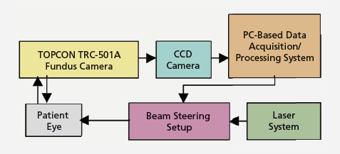
A major part of this system is the tracking of the retina using a computer-controlled system that captures and processes retinal images. Novel image processing routines are applied to the retinal images to determine the correct positions for laser shots. An accurate segmentation procedure is applied to the first retinal image to extract the sensitive areas in the retina. Subsequently, this segmentation is continuously updated using a fast registration procedure to obtain the new positions in case of eye movement at real-time rates. These positions can be fed to a beam-steering apparatus that precisely directs the laser beam accordingly.
The retinal images are acquired using a fundus camera. Contour detection is performed using deformable models. This technique is an iterative process in which one initiates an active contour model close to the contour of interest. The contour model is deformed using one of many algorithms for searching the neighboring points of each snaxel. All the points having a value above a certain threshold are extracted as new points on the contour. This not only deforms the contour but also makes it grow.
After extracting all sensitive objects that must be avoided during laser treatment, a binary image was composed containing these objects. This image was dilated by a square structuring element of dimensions 7×7 to maintain a safety margin around these sensitive areas. These locations are stored in the patient database, and get updated with every successive image frame.
A fast registration technique is proposed to automatically track the motion of segmented areas within the subsequent images in the sequence. In this technique, points satisfying certain developed significance conditions are chosen as landmarks in the reference frame. Using the extracted set of corresponding points in the subsequent image frames, one can accurately align image frames to compensate for eye movements and saccades.
This work was done by Nahed H. Solouma, Abou-Bakr M. Youssef, Yehia A. Badr, and Yasser M. Kadah of Biomedical Engineering Department and Laser Institute at Cairo University for the Army Research Laboratory. ARL-0064
This Brief includes a Technical Support Package (TSP).
Computer-Assisted Laser Treatment Using Real-Time Retinal Tracking
(reference ARL-0064) is currently available for download from the TSP library.
Don't have an account?
Overview
The document presents a novel computerized system aimed at improving laser treatment for choroidal neovascularization (CNV) associated with diabetic retinopathy, a leading cause of blindness. The system integrates a TOPCON TRC-501A fundus camera with a computer to facilitate real-time capturing and processing of retinal images. This approach allows for accurate treatment planning and execution, addressing the challenges faced in traditional laser treatments, which often require multiple sessions and have a low success rate.
The core of the system involves an advanced segmentation technique that extracts sensitive areas of the retina, including the blood vessel tree. This segmentation is crucial for identifying the boundaries of both wide and narrow blood vessels, ensuring that laser shots are accurately targeted while avoiding critical structures like the optic disc and macula. The segmentation process is designed to be as automatic as possible, minimizing user interaction and computational complexity.
To enhance the system's effectiveness, a fast registration technique is employed to track the motion of the retina in real-time. This technique uses a reference image to establish landmarks, which are then matched in subsequent images to compensate for eye movements and saccades. By aligning the images accurately, the system can direct laser shots precisely to the intended locations, thereby improving treatment outcomes.
The document highlights the computational aspects of the proposed algorithm, noting that the segmentation of the blood vessel tree is the most computationally intensive part, typically taking between 4-7 seconds per image. However, this processing time is manageable as it occurs before the actual laser treatment, allowing for real-time adjustments during the procedure.
Overall, the proposed system aims to significantly enhance the accuracy and efficiency of laser treatments for CNV, potentially increasing the success rates and reducing the recurrence of the condition. By leveraging advanced image processing techniques and real-time tracking, the system represents a significant advancement in the field of retinal laser therapy, promising better outcomes for patients suffering from diabetic retinopathy.