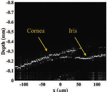
A new bioimaging device can operate with significantly lower power and in an entirely non-mechanical way. It could one day improve detecting eye and even heart conditions. The device uses a process called electrowetting to change the surface shape of a liquid to perform optical functions. By creating a device that doesn’t use scanning mirrors, the technique requires less electrical power than other devices used for OCT and bioimaging. To test the device’s ability to perform biomedical imaging, the researchers turned to zebrafish. The researchers focused on identifying where the cornea, iris, and retina was from the zebrafish. The two benchmarks that the group hoped to achieve were 10 μm in axial resolution and then around 5 μm in lateral resolution.
This device could open new doors for mapping aspects of the retina that can be essential for diagnosing potential eye conditions like age-related macular degeneration and glaucoma. The team hopes to create endoscopes that could revolutionize bioimaging technology. With these components, they can maintain a very small-scale optical system compared to a mechanical scanner that can help OCT technologies. (Image credit: EPFL/Alain Herzog)
For more information, visit here .