Researchers have developed a prototype imaging system that could significantly improve doctors’ ability to detect cancerous tissue during endoscopic procedures. This approach combines light-emitting diodes (LEDs) with hyperspectral imaging technology to create detailed maps of tissue properties that are invisible to conventional endoscopic cameras.
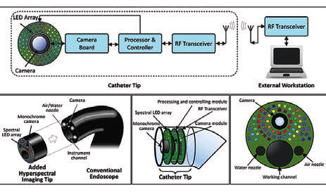
The team designed and tested a prototype system built around an array of 18 LEDs, each emitting light at different wavelengths ranging from 405 to 910 nm. The system uses a monochrome camera to capture images as each LED illuminates the target tissue in sequence, building up a complete hyperspectral dataset.
The researchers evaluated their LED-based system by imaging both normal and cancerous tissue samples removed during surgery. They investigated how different imaging conditions affected the quality of the hyperspectral data and compared their results with those from a reference hyperspectral camera system used as a gold standard. The researchers designed their system using wavelength scanning, where the LEDs illuminate the tissue sequentially at different wavelengths. (Image credit: N. Modir et al., doi 10.1117/1.JMI.12.3.035002)
For more information, visit here .