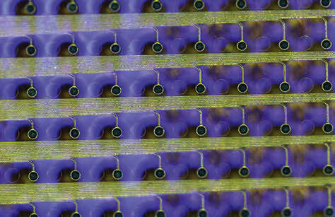
A thin film that combines an electrode grid and LEDs can both track and produce a visual representation of the brain’s activity in real time. The device is designed to provide neurosurgeons visual information about a patient’s brain to monitor brain states during surgical interventions to remove brain lesions including tumors and epileptic tissue.
Each LED in the device mirrors the activity of a few thousand neurons. In a series of proof-of-concept experiments in rodents and large non-primate mammals, researchers showed that the device can effectively track and display neural activity in the brain corresponding to different areas of the body. In this case, the LEDs developed by the team light up red in the areas that need to be removed by the surgeon. Surrounding areas that control critical functions and should be avoided show up in green.
The team used gallium nitride to develop a manufacturing technique for high-efficiency LEDs that do not heat up when they light up and do not damage brain tissues. The embedded thousands of LEDs in flexible films and release them from the substrate in the form of a flexible display panel. They then used inkjet printing to deposit quantum dot inks on the surface of the LEDs to convert their blue light to multiple other colors. (Image credit: UCSD)
For more information, visit here .

