A noninvasive imaging system combines two advanced techniques to examine both the structure and chemical composition of skin cancers. This approach could improve how doctors diagnose and classify skin cancer and how they monitor treatment responses.
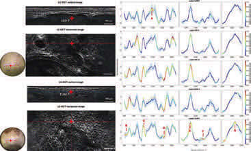
The system merges two types of imaging: line-field confocal optical coherence tomography (LC-OCT), which captures high-resolution images of skin tissue at the cellular level, and confocal Raman microspectroscopy, which analyzes the chemical makeup of specific areas identified in those images. Together, these tools allow researchers to not only see the shape and structure of cancerous cells but also understand their molecular characteristics.
The AI model performed well, achieving a classification accuracy of 95 percent for basal cell carcinoma and 92 percent when both types of cancer were included. These results suggest the system can reliably distinguish cancerous structures based on their chemical signatures. Further analysis revealed distinct chemical differences between various cancer types, offering new insights into how these cancers develop and behave. This dual-imaging approach could lead to more precise, less-invasive skin cancer diagnostics in the future. (Image credit: M. Ayadh et al., doi 10.1117/1.JBO.30.7.076008)
For more information, visit here .