Researchers have developed a first-of-its-kind wearable device capable of continuously scanning the lungs and heart of hospital patients while they rest in bed — offering a revolutionary alternative to CT scans.
The belt-like device, attached around a patient’s chest, uses ultrasound and works like a CT scanner. Rather than taking an isolated snapshot, it can produce a series of dynamic, high-resolution images of the heart, lungs and internal organs over time, giving doctors deeper insight into a patient’s condition. The device can be worn in bed and also reduces the need for repeated trips to radiology or exposure to doses of ionizing radiation.
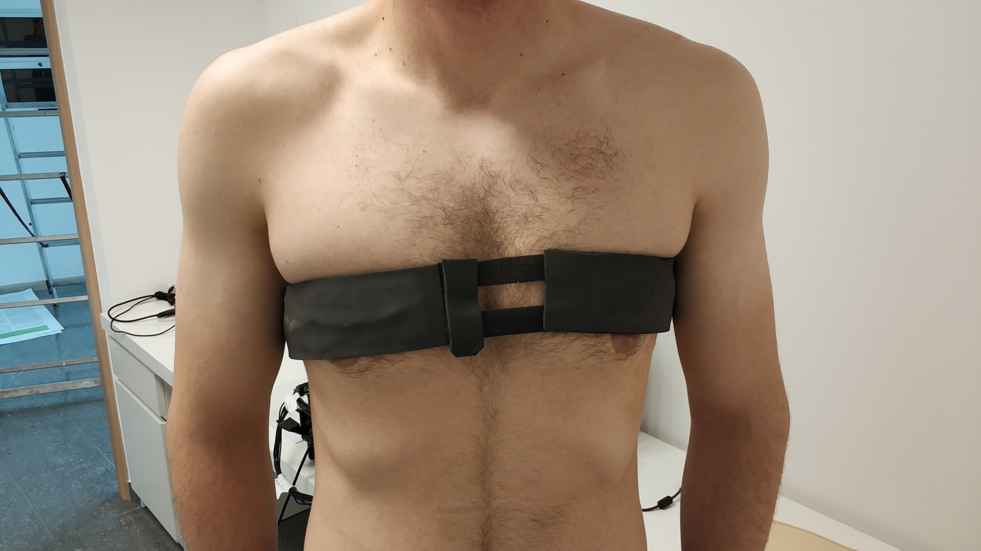
The soft, skin-conforming sensor array is placed directly on a patient's chest and uses sophisticated ultrasound computed tomography (USCT) to generate images of the heart and lungs in real time, tracking changes in organ function and structure continuously over hours or even days.
Crucially, the device is designed with patient comfort in mind. Its soft, flexible materials make it suitable for long-term wear, and its wireless data transmission capabilities allow integration with hospital monitoring systems. Future iterations may even offer AI-assisted analysis for clinicians, identifying warning signs before they’re visible to the human eye.
Beyond hospitals, this technology opens the door to remote monitoring in home care settings, particularly for elderly patients or those with chronic cardiopulmonary diseases. It may also reduce the healthcare burden by preventing unnecessary hospital admissions through early intervention.