Besides radiotherapy and chemotherapy, the standard procedure for treating brain tumors includes the surgical removal of all or some of the growth. This surgery calls for precision and perfection — both from the surgeon and the instruments, because knowledge and skill alone do not determine the successful outcome of surgery.
Time is also a factor. Constantly stopping to adjust the microscope interrupts the procedure and consumes valuable minutes when the surgeon’s hands could be completing the procedure. As a result, the dose of anesthetics must be increased, and this puts the patient under additional stress. The loss of time can even be a matter of life and death.
Solving this problem became a goal of David Pitskhelauri, MD, Ph.D., a brain tumor and surgical epilepsy therapy specialist at the Moscow Burdenko Neurosurgery Institute in Moscow, Russia. He knew that if the microscope had a hands-free setting, the surgeon would be able to operate without stopping. And so the doctor, along with a product development firm, a manufacturer, and a specialist in electrical connector technology joined forces to develop Mari, a precision instrument that enables the hands-free use of a surgical microscope, heralding a new age of neurosurgery.
From Concept to Development
Help in creating the solution came from product development firm Astratech, also based in Moscow. The firm’s director, Nicolay Rozhnin, Ph.D., led the development of the system, which reacts to the slightest movements of the surgeon’s lips and head and converts them into control signals. Sensitivity, precision, and quick reactions were all essential. In addition to Dr. Pitskhelauri, who addressed the medical requirements, the development team included Astratech experts in electronics and microtechnology and connector technology company ODU. Tolikety Co. was selected to manufacture the Mari microscope attachment.
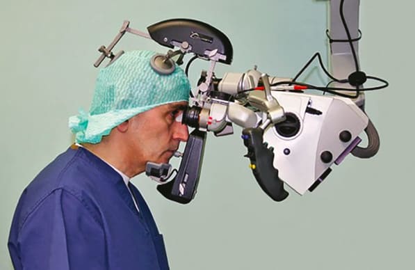
What exactly is Mari? The Mari precision control system is used in conjunction with a multifunction counterweight-balanced surgical microscope.1 It consists of a rectangular titanium holder with supportive plates, a frame that allows the device to be attached to the microscope eyepieces, a joystick, and an electric switch for control of the microscope’s functions. A microprocessor, a small electric drive with a locking mechanism, an electric heating system, and a multiwire cable provide the rest of the functionality. A 12-V transformer is used for the power supply.
The Mari device resembles a helmet and is relatively heavy at 1100 g (2.43 lb). The bulk of the weight is carried by the microscope used in the surgery, keeping the weight of the device off of the surgeon. The device is attached to the surgeon’s head in such a way that the surgeon feels only mild contact with the support plates with no excessive pressure on the head.
The electric switch in the Mari chin rest releases the magnetic supports for the microscope so that it can be moved and turned along the x, y, z axes as well as on the rotational and diagonal axes. The joystick is positioned at the height of the surgeon’s mouth and is moved by the surgeon’s lips to adjust the microscope focus and zoom. The signal transmission to the surgical microscope, including focus-up, focus-down, zoom-in, and zoom-out, are also regulated by the surgeon’s lips.
The Key Role of Connectors
Connections for medical applications must meet the most exacting customer specifications and market standards for precision, reliability, and product safety. Health, correct diagnosis, and sometimes even life can depend on the fault-free operation of connectors. The Mari device with its precision control system is a prime example of the need for connectors that can ensure that the device meets these stringent requirements.2
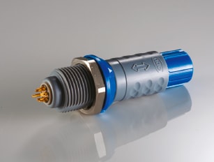
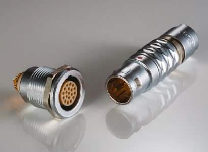
For Mari, the customized, application-specific ODU connectors together with the cable assemblies are key components of the overall system, ensuring reliable power and signal transmission between the control unit and the monitoring system of the surgical microscope. Not only does the system contain ODU’s connectors on each side of the Mari device, but additional ODU Mini-Snap push-pull connectors with a total of six different cable assemblies are used within the precision control system. The highly reliable circular ODU Mini-Snap connectors serve a dual purpose of supplying the control unit and small electromagnets with power and for ensuring an uninterrupted signal transmission to the microscope.
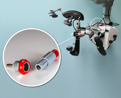
A multiwire cable provides a connection between the Mari device and the surgical microscope control system. The cable is connected to the microscope via a multiplug socket designed for a foot control panel. The device frame and heating system are attached to the binoculars. A push-pull cable assembly is used to heat the oculars on the control unit to prevent them from misting over — and therefore obstructing the surgeon’s view — during the surgery. The heating system generates a temperature up to 30–35 °C and prevents fogging of the surface of the eyepieces.
The pushing mechanism in the Mari device is fastened to the left handgrip and is connected to a multiwire cable. With the push-pull locking system, the connection secures itself when mated, and the push-pull locking principle ensures that there is no accidental disconnection. When the operator is ready, the plug can easily be demated from the receptacle by pulling back on the outer sleeve of the connector housing.
The ODU Mini-Snap push-pull locking connector technology offers multiple keying options and IP50 and IP68 protection. The connectors, which facilitate blind mating, provide reliable connections for more than 5,000 mating cycles. In addition to coax cable, triax cable is also available for critical applications. Triax offers an extra layer of insulation and a second conducting sheath, and it provides greater bandwidth as well as greater immunity to interference than coax. Other options include color coding, metal or plastic housings, and versions with the potential for autoclave and sterilizer use. The customizable RoHS-compliant connectors come in solder, crimp, and PCB termination types, and in 2-40 pin insert configurations.
Conclusion
The Mari device has been particularly well received in the field of neurosurgery. It is being used successfully in Russia as well as in Europe. The device is patented in the United States, the Russian Federation, and the European Union, and Mari has been approved for use in Russia, the European Union, and the United States. There were more than 2500 successful operations, including in-depth, hard-to-reach neoplasms, with Mari at the Burdenko Neurosurgery Institute.
The development of the Mari precision control system has the potential to change the way surgeons approach neurosurgery. Constantly stopping to adjust the microscope will be a thing of the past because the surgeon’s hands can be free to complete the procedure. For Mari, the customized, application-specific ODU connectors and cable assemblies were critical to ensuring reliable power and signal transmission between the control unit and the monitoring system of the surgical microscope.
References
- D. I. Pitskhelauri et al., “A Novel Device for Hands-Free Positioning and Adjustment of the Surgical Microscope,” J Neurosurg, p. 121:161–164, 2014; published online April 25, 2014; DOI: 10.3171/2014.3.JNS12578.
- M. Samoylova, “Mari,” Der Steckverbinder, p. 14–17, 2015.
This article was written by Marina Samoylova, ODU representative in Russia, CIS, Latvia, and Lithuania, and Anne Meimeth, Uschi vogg PR, Munich, Germany. For more information, click here .
A video on the Mari technology can be viewed at www.youtube.com/watch?v=BcpUXuIQx3I .
Transcript
00:00:00 the operating microscope is one of the most vital pieces of equipment in microsurgical operating rooms in particularly neurosurgical one modern counterweight balance microscope can be navigated in all required planes of movements with the help of surgeons hands in order to overcome the necessity of manual control over the microscope and to facilitate microsurgical
00:00:20 manipulations we propose the mark device which provides hands-free positioning of the operating microscope and adjustment of its optic system this device is designed from the navigation of the counterweight balanced microscope and consists of the rigid titanium part with supportive plates a locking mechanism a joystick and an electric switch located within the chin of the supported played
00:00:49 a special fixated frame is easily attached to the Beinecke or tip of the microscope the device is fixed to the frame with a single screw multi-channel cable provides a connection between the microscope and the device the fixative frame located on the binocular tube does not cause any inconvenience for surgeon who operates without the device the electric heating
00:01:31 system which prevents fogging on the surface of the eyepieces is attached to the binoculars you the electric switch in the chin supported plate of the device provides the release I know for the magnetic clutches of the Marksville when the switch is pressed the pushing mechanisms on the hands grip is activated that is
00:02:13 the release of all joints of the Marc's hope to begin the microsurgical manipulations the surgeon takes the required position in relation to the microscope and by pressing the joystick with his or her lips locks the device to navigate the microscope in the desired direction the surgeon slightly opens his or her mouth and moves his or her chin forward
00:02:36 opening the mouth and chin moment leads to tight connection of the surgeon's head with the device due to the increased distance between the paradoxical and Chi regions and pressing the electrical switch for eyes the release of all magnetic joys of the microscope now the surgeon can navigate the microscope with little effort in all directions including not only
00:02:53 translational moments along XYZ axes but also rotation ones which gives an advantage over the amount which device for microscope navigation the surgeon by closing his or her mouth simultaneously releases the electrical switch of the device it locks magnetic patches of the microscope this is how the microscope is navigated by the surgeon
00:03:24 you by pressing the joystick the surgeon can unlock the device and release his or her head the device permits hands-free control over the microscopes optic system the multifunctional joystick provides not only closing and opening of the device but also adjustment of focus and zoom slight motions of the surgeons leaves to the right to the left
00:03:53 upwards and downwards provide the transmission of corresponding signal focus up focus down zoom in and zoom out to the microscope control system the murid device provides very precise movements of the microscope unless the surgeon to manipulate without taking his or her hands out of operating field this is the demonstration of the virtual surgical procedure where the surgeon
00:04:15 tries to block the count with small features you supporter played in the nosebleed area provide static position of the surgeon's eyes floated to the binoculars so that the permanent view of the operative field is sustained during the microscope moment you
00:04:49 advantage of the Martin device is obvious it is clear that without my device all these manipulations would have been much more complicated the devices attached to the stereomicroscope without any difficulties or parts involving fixated frames cables heating system except metallic holder are located under the sterile drape the cable with an electric
00:05:19 plug is pulled out of the sterile drape through a small opening near the binder course and connected to the device the mark device provides easy and accurate navigation of the operation microscope in great range of moments around the operated field the object parameters can be adjusted quickly when changing the position of the microscope it is important to note that surgeons
00:05:38 head is not compressed when the microscope is kept in static position the surgeon feels only a mild contact with the supporting plates which causes no discomfort this video represents the course of surgical removal of the left optic nerve hemangioblastoma without using any rigid retractors the surgeon frequently changes his position around the craniotomy side in order to use all
00:05:59 available exposure without disrupting surgical procedure the mark device significantly facilitates microsurgical manipulations the optic parameters can be adjusted easily and quickly when changing the position of the microscope the device allows the surgeon to perform surgical manipulations through the deep and narrow operator corridor without using brain retractors Mauri device
00:06:22 allows to increase an accuracy of the surgical manipulations and to shorten the duration of the surgery you when needed by pressing the joystick the surgeon can unlock the device and release his or her head the inventor of the device has applied it in about 600 operations raging in difficulty and with duration up to 7
00:07:03 hours it is possible to adjust the model device to all types of the counterweight bronze microscopes the device can be useful in all fields of surgery where operating microscope is required the device is packed in a small suitcase and can be easily transported