No, “CSI: Ocean” is not the next installment of the television franchise that investigates crime scenes. Nevertheless, one group of scientists and engineers combine their access to specialized tools, ships, labs, and underwater vehicles with skill and knowledge to create a detailed understanding of the global ocean system and its inhabitants.

As part of its mission, the Woods Hole Oceanographic Institution, in Woods Hole, MA, studies sea animals that have washed up to help determine what natural or man-made effects are responsible for their deaths, including a recent dolphin die-off, working with their own CSI group: the Computerized Scanning and Imaging Facility.
A sophisticated CT (computer tomography) scanner is used to obtain high-resolution mappings of the various aquatic animals, not just to determine cause of death, but also to understand how they survive in the ocean. Of special interest is understanding more on how echolocation operates by studying auditory anatomy. They are also studying how dolphins can dive deeply and surface quickly, normally without suffering from “the bends,” nitrogen bubbles forming in the blood stream.
CT is invaluable for understanding internal structure. Two- and three-dimensional visualizations provide valuable data for research topics as diverse as tissue and sound interactions, climate change, reef structure, and core analyses. Researchers interested in functional anatomy or pathology now have the opportunity to visualize tissues in situ and undisturbed. This includes fine structures of soft tissue, fluids, fats, air, and bone.
CSI’s CT scanner can image a wide range of densities of varying materials and specimens at an optimal resolution of 100 micron voxels or, for rapid imaging, at table speeds up to 1m/min data acquisition. In practical terms, this means data showing internal differences in tissue density of a 100 to 200 kg, two meter dolphin can be acquired in just over two minutes and imaged at 100 micron intervals through the entire body. X-ray attenuations can be measured up to 40,000 gradations or Hounsfield units.
The high-resolution scans needed for these and other studies require precise positioning and movement of the animal being studied in a CT scanner. For some studies, the deceased animal is suspended in water under various pressures to mimic dive gradients. This combination of the animal and the surrounding water can be on the order of 3,100 lbs (1,400 kg), much higher than the standard tables designed to position people within the scanner. Any positioning error, including vibrations, distort the images and may make the image less useful for resolving the questions of the investigation.
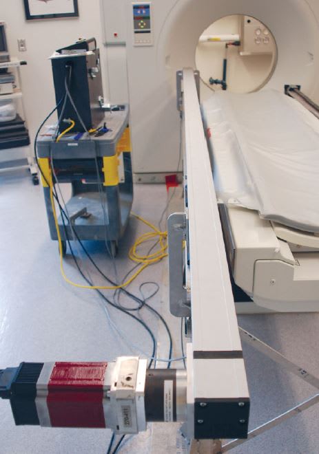
A custom scan drive was designed by Artec Imaging to allow motion on the range of 0.5 to 10 mm per second allowing slice reconstruction down to 0.1mm. The table is synchronized to the motion table normally used to position patients. This allows for positioning of heavy loads without needing to modify the scanners.
A QCI-A34HC-2 motor from QuickSilver Controls, Inc., is used to move the transport table. (See Figure 1) The high torque capability of the motor combined with the high inertial mismatch capability of the controller allows the table to use the same tuning parameters loaded or unloaded. The resulting motions are very smooth, as needed for accurate scans, while still providing the capability for rapid motions allowing quick positioning of the specimen prior to scanning.
This article was written by Don Labriola, PE, President, QuickSilver Controls, Inc., Covina, CA. For more information on the CSI at Woods Hole, visit http://csi.whoi.edu/ . For more information on Artec Imaging, Cornelius, NC, visit http://info.hotims.com/49741-167 . For more information on QuickSilver Controls, visit http://info.hotims.com/49741-166 .


